
ELK Biotechnology
SKU:ES6787
PKD1 (phospho Tyr463) Rabbit Polyclonal Antibody
PKD1 (phospho Tyr463) Rabbit Polyclonal Antibody
Regular price
$248.00 USD
Regular price
Sale price
$248.00 USD
Unit price
per
Shipping calculated at checkout.
Couldn't load pickup availability
PKD1 (phospho Tyr463) Rabbit Polyclonal Antibody
Overview
-
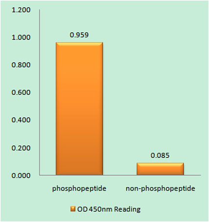 Enzyme-Linked Immunosorbent Assay (Phospho-ELISA) for Immunogen Phosphopeptide (Phospho-left) and Non-Phosphopeptide (Phospho-right), using PKD1/PKC mu (Phospho-Tyr463) Antibody
 -
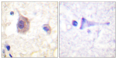 Immunohistochemistry analysis of paraffin-embedded human brain, using PKD1/PKC mu (Phospho-Tyr463) Antibody. The picture on the right is blocked with the phospho peptide.
 -
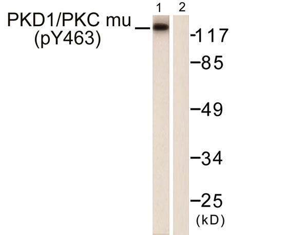 Western blot analysis of lysates from HepG2 cells, using PKD1/PKC mu (Phospho-Tyr463) Antibody. The lane on the right is blocked with the phospho peptide.
