1
/
of
1
ELK Biotechnology
SKU:ES6572
Peroxin 7 Rabbit Polyclonal Antibody
Regular price
$248.00 USD
Regular price
Sale price
$248.00 USD
Unit price
/
per
Shipping calculated at checkout.
Couldn't load pickup availability
Peroxin 7 Rabbit Polyclonal Antibody
Overview
| Product name: | Peroxin 7 rabbit pAb |
| Alternative Names: | PEX7; PTS2R; Peroxisomal targeting signal 2 receptor; PTS2 receptor; Peroxin-7 |
| Applications: | WB;IHC;IF;ELISA |
| Recommended Dilutions: | Western Blot: 1/500 - 1/2000. Immunohistochemistry: 1/100 - 1/300. ELISA: 1/40000. Not yet tested in other applications. |
| Immunogen: | The antiserum was produced against synthesized peptide derived from human PEX7. AA range:204-253 |
| Storage: | Rabbit |
| Storage: | -20°C/1 year |
| Clonality: | Polyclonal |
| Isotype: | IgG |
| Concentration: | 1 mg/ml |
| Observed Band: | 40kD |
| GeneID(Human): | 5191 |
| Human Swiss-Prot No: | O00628 |
| Cellular localization: | Peroxisome . Cytoplasm . |
| Background: | This gene encodes the cytosolic receptor for the set of peroxisomal matrix enzymes targeted to the organelle by the peroxisome targeting signal 2 (PTS2). Defects in this gene cause peroxisome biogenesis disorders (PBDs), which are characterized by multiple defects in peroxisome function. There are at least 14 complementation groups for PBDs, with more than one phenotype being observed in cases falling into particular complementation groups. Although the clinical features of PBD patients vary, cells from all PBD patients exhibit a defect in the import of one or more classes of peroxisomal matrix proteins into the organelle. Defects in this gene have been associated with PBD complementation group 11 (PBD-CG11) disorders, rhizomelic chondrodysplasia punctata type 1 (RCDP1), and Refsum disease (RD). [provided by RefSeq, Oct 2008], |
| Species Reactivity: | Human;Mouse;Rat |
-
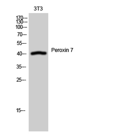
Western Blot analysis of 3T3 cells using Peroxin 7 Polyclonal Antibody -
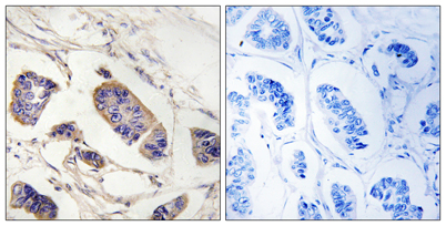
Immunohistochemistry analysis of paraffin-embedded human breast carcinoma tissue, using PEX7 Antibody. The picture on the right is blocked with the synthesized peptide. -
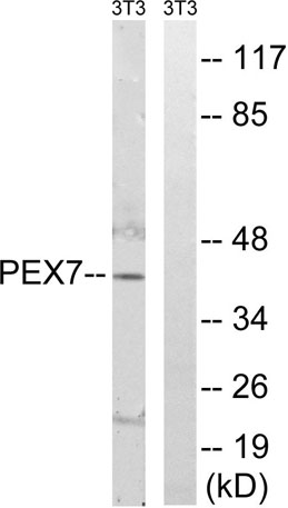
Western blot analysis of lysates from NIH/3T3 cells, using PEX7 Antibody. The lane on the right is blocked with the synthesized peptide.
Share
