MRPS16; RPMS16; CGI-132; 28S ribosomal protein S16; mitochondrial; MRP-S16; S16mt
Applications:
WB;IHC;IF;ELISA
Recommended Dilutions:
Western Blot: 1/500 - 1/2000. Immunohistochemistry: 1/100 - 1/300. ELISA: 1/40000. Not yet tested in other applications.
Immunogen:
The antiserum was produced against synthesized peptide derived from human MRPS16. AA range:81-130
Storage:
Rabbit
Storage:
-20°C/1 year
Clonality:
Polyclonal
Isotype:
IgG
Concentration:
1 mg/ml
Observed Band:
15kD
GeneID(Human):
51021
Human Swiss-Prot No:
Q9Y3D3
Cellular localization:
Mitochondrion .
Background:
Mammalian mitochondrial ribosomal proteins are encoded by nuclear genes and help in protein synthesis within the mitochondrion. Mitochondrial ribosomes (mitoribosomes) consist of a small 28S subunit and a large 39S subunit. They have an estimated 75% protein to rRNA composition compared to prokaryotic ribosomes, where this ratio is reversed. Another difference between mammalian mitoribosomes and prokaryotic ribosomes is that the latter contain a 5S rRNA. Among different species, the proteins comprising the mitoribosome differ greatly in sequence, and sometimes in biochemical properties, which prevents easy recognition by sequence homology. This gene encodes a 28S subunit protein that belongs to the ribosomal protein S16P family. The encoded protein is one of the most highly conserved ribosomal proteins between mammalian and yeast mitochondria. Three pseudogenes (located at 8q21.3, 20

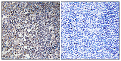 Immunohistochemistry analysis of paraffin-embedded human tonsil tissue, using MRPS16 Antibody. The picture on the right is blocked with the synthesized peptide.
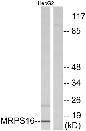 Western blot analysis of lysates from HepG2 cells, using MRPS16 Antibody. The lane on the right is blocked with the synthesized peptide.
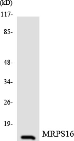 Western blot analysis of the lysates from COLO205 cells using MRPS16 antibody.