Western Blot: 1/500 - 1/2000. Immunohistochemistry: 1/100 - 1/300. ELISA: 1/40000. Not yet tested in other applications.
Immunogen:
The antiserum was produced against synthesized peptide derived from human NDUFS1. AA range:620-669
Storage:
Rabbit
Storage:
-20°C/1 year
Clonality:
Polyclonal
Isotype:
IgG
Concentration:
1 mg/ml
Observed Band:
80kD
GeneID(Human):
4719
Human Swiss-Prot No:
P28331
Cellular localization:
Mitochondrion inner membrane ; Peripheral membrane protein ; Matrix side .
Background:
The protein encoded by this gene belongs to the complex I 75 kDa subunit family. Mammalian complex I is composed of 45 different subunits. It locates at the mitochondrial inner membrane. This protein has NADH dehydrogenase activity and oxidoreductase activity. It transfers electrons from NADH to the respiratory chain. The immediate electron acceptor for the enzyme is believed to be ubiquinone. This protein is the largest subunit of complex I and it is a component of the iron-sulfur (IP) fragment of the enzyme. It may form part of the active site crevice where NADH is oxidized. Mutations in this gene are associated with complex I deficiency. Several transcript variants encoding different isoforms have been found for this gene. [provided by RefSeq, Jan 2011],

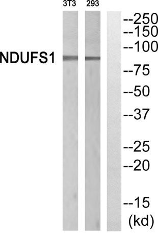 Western blot analysis of NDUFS1 Antibody. The lane on the right is blocked with the NDUFS1 peptide.
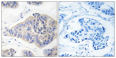 Immunohistochemistryt analysis of paraffin-embedded human breast carcinoma, using NDUFS1 Antibody. The lane on the right is blocked with the NDUFS1 peptide.
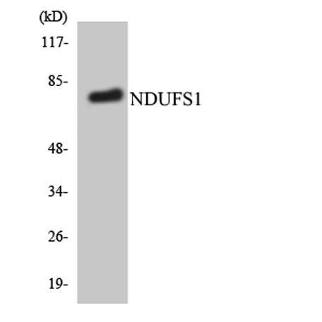 Western blot analysis of the lysates from HepG2 cells using NDUFS1 antibody.