ATP5L2; ATP5K2; ATP synthase subunit g 2; mitochondrial; ATPase subunit g 2
Applications:
WB;IF;ELISA
Recommended Dilutions:
Western Blot: 1/500 - 1/2000. Immunofluorescence: 1/200 - 1/1000. ELISA: 1/40000. Not yet tested in other applications.
Immunogen:
The antiserum was produced against synthesized peptide derived from human ATP5L2. AA range:51-100
Storage:
Rabbit
Storage:
-20°C/1 year
Clonality:
Polyclonal
Isotype:
IgG
Concentration:
1 mg/ml
Observed Band:
20kD
GeneID(Human):
267020
Human Swiss-Prot No:
Q7Z4Y8
Cellular localization:
Mitochondrion membrane .
Background:
function:Mitochondrial membrane ATP synthase (F(1)F(0) ATP synthase or Complex V) produces ATP from ADP in the presence of a proton gradient across the membrane which is generated by electron transport complexes of the respiratory chain. F-type ATPases consist of two structural domains, F(1) - containing the extramembraneous catalytic core, and F(0) - containing the membrane proton channel, linked together by a central stalk and a peripheral stalk. During catalysis, ATP synthesis in the catalytic domain of F(1) is coupled via a rotary mechanism of the central stalk subunits to proton translocation. Part of the complex F(0) domain. Minor subunit located with subunit a in the membrane.,similarity:Belongs to the ATPase g subunit family.,subunit:F-type ATPases have 2 components, CF(1) - the catalytic core - and CF(0) - the membrane proton channel. CF(0) seems to have nine subunits: a, b, c, d, e, f, g, F6 and 8 (or A6L).,

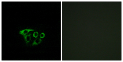 Immunofluorescence analysis of A549 cells, using ATP5L2 Antibody. The picture on the right is blocked with the synthesized peptide.
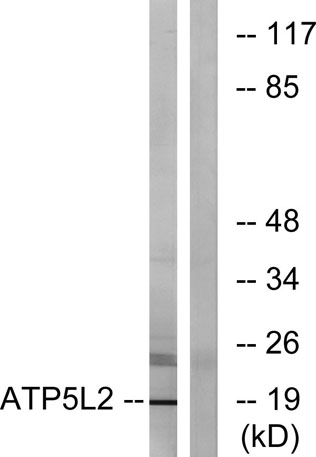 Western blot analysis of lysates from A549 cells, using ATP5L2 Antibody. The lane on the right is blocked with the synthesized peptide.
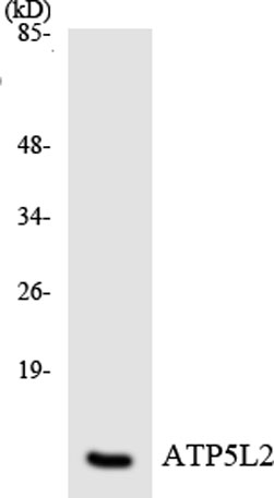 Western blot analysis of the lysates from HeLa cells using ATP5L2 antibody.