APOBEC3A; Probable DNA dC->dU-editing enzyme APOBEC-3A; Phorbolin-1
Applications:
WB;ELISA
Recommended Dilutions:
Western Blot: 1/500 - 1/2000. ELISA: 1/10000. Not yet tested in other applications.
Immunogen:
The antiserum was produced against synthesized peptide derived from human APOBEC3A. AA range:27-76
Storage:
Rabbit
Storage:
-20°C/1 year
Clonality:
Polyclonal
Isotype:
IgG
Concentration:
1 mg/ml
Observed Band:
26kD
GeneID(Human):
200315
Human Swiss-Prot No:
P31941
Cellular localization:
Nucleus. Cytoplasm.
Background:
This gene is a member of the cytidine deaminase gene family. It is one of seven related genes or pseudogenes found in a cluster, thought to result from gene duplication, on chromosome 22. Members of the cluster encode proteins that are structurally and functionally related to the C to U RNA-editing cytidine deaminase APOBEC1. The protein encoded by this gene lacks the zinc binding activity of other family members. The protein plays a role in immunity, by restricting transmission of foreign DNA such as viruses. One mechanism of foreign DNA restriction is deamination of foreign double-stranded DNA cytidines to uridines, which leads to DNA degradation. However, other mechanisms are also thought to be involved, as anti-viral effect is not dependent on deaminase activity. Two transcript variants encoding different isoforms have been found for this gene. [provided b

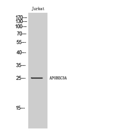 Western Blot analysis of Jurkat cells using APOBEC3A Polyclonal Antibody
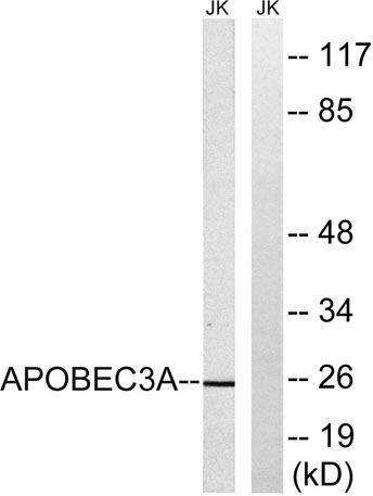 Western blot analysis of lysates from Jurkat cells, using APOBEC3A Antibody. The lane on the right is blocked with the synthesized peptide.
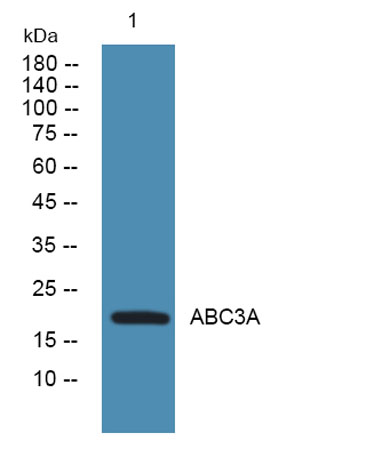 Western blot analysis of lysates from K562 cells, primary antibody was diluted at 1:1000, 4°over night