
ELK Biotechnology
SKU:ES4402
P-cadherin Rabbit Polyclonal Antibody
P-cadherin Rabbit Polyclonal Antibody
Regular price
$248.00 USD
Regular price
Sale price
$248.00 USD
Unit price
per
Shipping calculated at checkout.
Couldn't load pickup availability
P-cadherin Rabbit Polyclonal Antibody
Overview
-
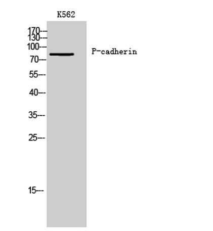 Western Blot analysis of K562 cells using P-cadherin Polyclonal Antibody
 -
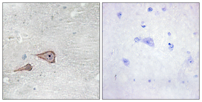 Immunohistochemistry analysis of paraffin-embedded human brain tissue, using CDH3 Antibody. The picture on the right is blocked with the synthesized peptide.
 -
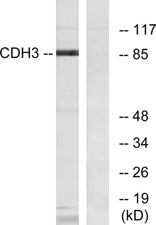 Western blot analysis of lysates from K562 cells, using CDH3 Antibody. The lane on the right is blocked with the synthesized peptide.
 -
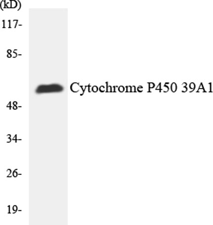 Western blot analysis of the lysates from HepG2 cells using Cytochrome P450 2C19 antibody.
