
ELK Biotechnology
SKU:ES3622
TNF-R2 Rabbit Polyclonal Antibody
TNF-R2 Rabbit Polyclonal Antibody
Regular price
$248.00 USD
Regular price
Sale price
$248.00 USD
Unit price
per
Shipping calculated at checkout.
Couldn't load pickup availability
TNF-R2 Rabbit Polyclonal Antibody
Overview
-
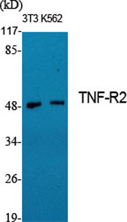 Western Blot analysis of various cells using TNF-R2 Polyclonal Antibody diluted at 1:1000. Secondary antibody(catalog#:RS0002) was diluted at 1:20000
 -
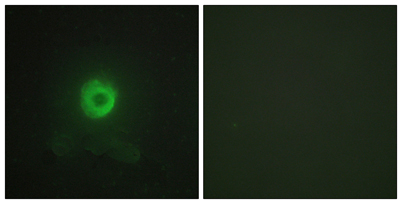 Immunofluorescence analysis of HeLa cells, using TNF Receptor II Antibody. The picture on the right is blocked with the synthesized peptide.
 -
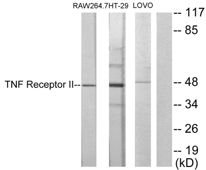 Western blot analysis of lysates from RAW264.7, HT-29, and LOVO cells, using TNF Receptor II Antibody. The lane on the right is blocked with the synthesized peptide.
 -
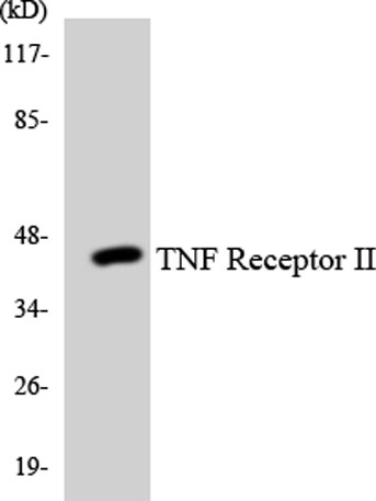 Western blot analysis of the lysates from HUVECcells using TNF Receptor II antibody.
