ELK Biotechnology
SKU:ES3328
RARβ Rabbit Polyclonal Antibody
RARβ Rabbit Polyclonal Antibody
Regular price
$248.00 USD
Regular price
Sale price
$248.00 USD
Unit price
per
Shipping calculated at checkout.
Couldn't load pickup availability
RARβ Rabbit Polyclonal Antibody
Overview
-
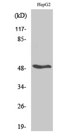 Western Blot analysis of various cells using RARβ Polyclonal Antibody
 -
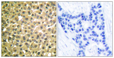 Immunohistochemistry analysis of paraffin-embedded human breast carcinoma tissue, using Retinoic Acid Receptor beta Antibody. The picture on the right is blocked with the synthesized peptide.
 -
 Western blot analysis of lysates from HepG2 cells, using Retinoic Acid Receptor beta Antibody. The lane on the right is blocked with the synthesized peptide.
 -
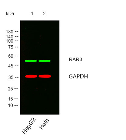 Western blot analysis of lysates from HepG2,Hela cells, (Green) primary antibody was diluted at 1:1000, 4°over night, secondary antibody was diluted at 1:10000, 37° 1hour. (Red) loading contrl antibody was diluted at 1:5000 as loading control, 4° over nig