ELK Biotechnology
SKU:ES3284
PU.1 Rabbit Polyclonal Antibody
PU.1 Rabbit Polyclonal Antibody
Regular price
$248.00 USD
Regular price
Sale price
$248.00 USD
Unit price
per
Shipping calculated at checkout.
Couldn't load pickup availability
PU.1 Rabbit Polyclonal Antibody
Overview
-
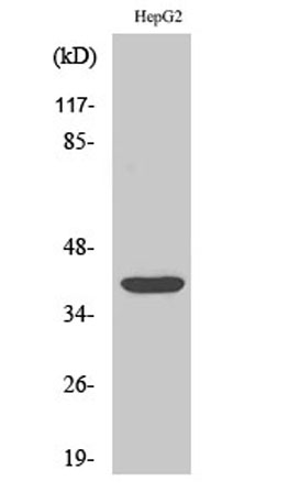 Western Blot analysis of various cells using PU.1 Polyclonal Antibody cells nucleus extracted by Minute TM Cytoplasmic and Nuclear Fractionation kit (SC-003,Inventbiotech,MN,USA).
 -
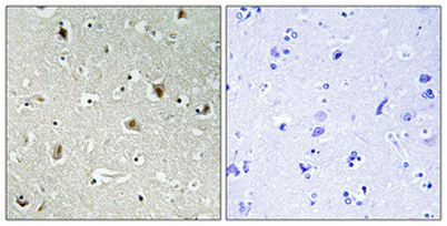 Immunohistochemical analysis of paraffin-embedded Human brain. Antibody was diluted at 1:100(4° overnight). High-pressure and temperature Tris-EDTA,pH8.0 was used for antigen retrieval. Negetive contrl (right) obtaned from antibody was pre-absorbed by i
 -
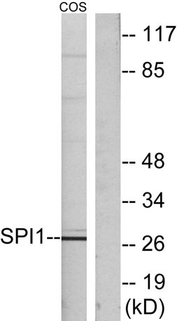 Western blot analysis of lysates from COS7 cells, using SPI1 Antibody. The lane on the right is blocked with the synthesized peptide.
 -
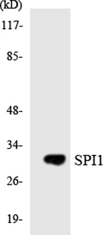 Western blot analysis of the lysates from HUVECcells using SPI1 antibody.