
ELK Biotechnology
SKU:ES2881
Myp Rabbit Polyclonal Antibody
Myp Rabbit Polyclonal Antibody
Regular price
$248.00 USD
Regular price
Sale price
$248.00 USD
Unit price
per
Shipping calculated at checkout.
Couldn't load pickup availability
Myp Rabbit Polyclonal Antibody
Overview
-
 Western Blot analysis of various cells using Myp Polyclonal Antibody
 -
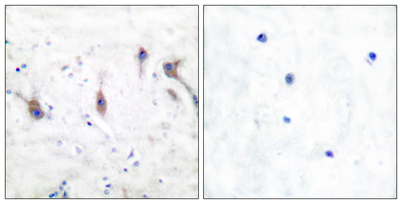 Immunohistochemistry analysis of paraffin-embedded human brain tissue, using ARC Antibody. The picture on the right is blocked with the synthesized peptide.
 -
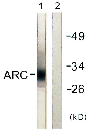 Western blot analysis of lysates from HeLa cells, using ARC Antibody. The lane on the right is blocked with the synthesized peptide.
 -
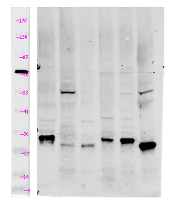 Western Blot analysis of lane1 mouse-brain, lane2 mouse-kidney, lane3 Hela. land4 MCF7, lane5 293T, lane6 mouse-muscle using primary antibody at 1:1000 dilution 4°C, overnight. Secondary antibody(catalog#:RS23920) was diluted at 1:10000 25°C,1.5hours
