ELK Biotechnology
SKU:ES2853
MRP-S36 Rabbit Polyclonal Antibody
MRP-S36 Rabbit Polyclonal Antibody
Regular price
$248.00 USD
Regular price
Sale price
$248.00 USD
Unit price
per
Shipping calculated at checkout.
Couldn't load pickup availability
MRP-S36 Rabbit Polyclonal Antibody
Overview
-
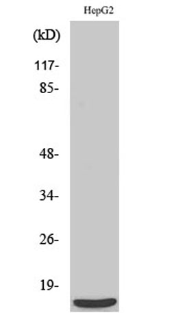 Western Blot analysis of various cells using MRP-S36 Polyclonal Antibody
 -
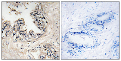 Immunohistochemistry analysis of paraffin-embedded human prostate carcinoma tissue, using MRPS36 Antibody. The picture on the right is blocked with the synthesized peptide.
 -
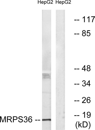 Western blot analysis of lysates from HepG2 cells, using MRPS36 Antibody. The lane on the right is blocked with the synthesized peptide.
 -
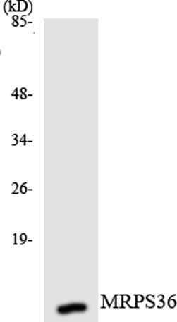 Western blot analysis of the lysates from K562 cells using MRPS36 antibody.