1
/
of
1
ELK Biotechnology
SKU:ES2747
MARK2 Rabbit Polyclonal Antibody
Regular price
$248.00 USD
Regular price
Sale price
$248.00 USD
Unit price
/
per
Shipping calculated at checkout.
Couldn't load pickup availability
MARK2 Rabbit Polyclonal Antibody
Overview
| Product name: | MARK2 rabbit pAb |
| Alternative Names: | MARK2; EMK1; Serine/threonine-protein kinase MARK2; ELKL motif kinase 1; EMK-1; MAP/microtubule affinity-regulating kinase 2; PAR1 homolog; PAR1 homolog b; Par-1b; Par1b |
| Applications: | WB;IF;ELISA |
| Recommended Dilutions: | Western Blot: 1/500 - 1/2000. Immunofluorescence: 1/200 - 1/1000. ELISA: 1/20000. Not yet tested in other applications. |
| Immunogen: | The antiserum was produced against synthesized peptide derived from human MARK2. AA range:10-59 |
| Storage: | Rabbit |
| Storage: | -20°C/1 year |
| Clonality: | Polyclonal |
| Isotype: | IgG |
| Concentration: | 1 mg/ml |
| Observed Band: | 85kD |
| GeneID(Human): | 2011 |
| Human Swiss-Prot No: | Q7KZI7 |
| Cellular localization: | Cell membrane; Peripheral membrane protein. Cytoplasm. Lateral cell membrane. Cytoplasm, cytoskeleton. Cell projection, dendrite . Cytoplasm . Phosphorylation at Thr-596 by PRKCZ/aPKC and subsequent interaction with 14-3-3 protein YWHAZ promotes relocation from the cell membrane to the cytoplasm. |
| Background: | microtubule affinity regulating kinase 2(MARK2) Homo sapiens This gene encodes a member of the Par-1 family of serine/threonine protein kinases. The protein is an important regulator of cell polarity in epithelial and neuronal cells, and also controls the stability of microtubules through phosphorylation and inactivation of several microtubule-associating proteins. The protein localizes to cell membranes. Multiple transcript variants encoding different isoforms have been found for this gene. [provided by RefSeq, Jul 2009], |
| Species Reactivity: | Human;Mouse;Rat;Monkey |
-
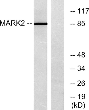
Western Blot analysis of various cells using MARK2 Polyclonal Antibody -
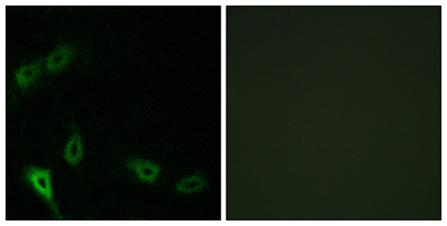
Immunofluorescence analysis of A549 cells, using MARK2 Antibody. The picture on the right is blocked with the synthesized peptide. -
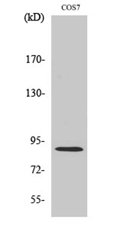
Western blot analysis of lysates from COS7 cells, using MARK2 Antibody. The lane on the right is blocked with the synthesized peptide. -
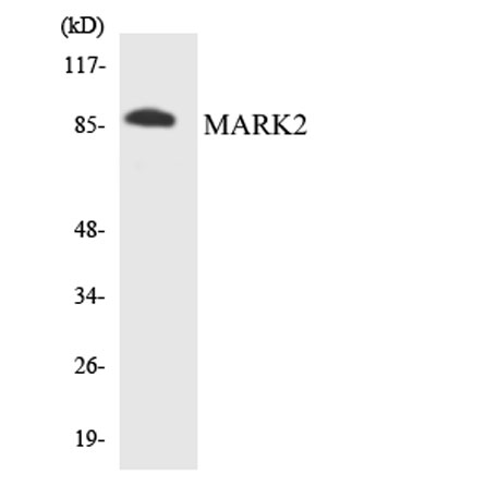
Western blot analysis of the lysates from 293 cells using MARK2 antibody.
Share
