Western Blot: 1/500 - 1/2000. Immunohistochemistry: 1/100 - 1/300. Immunofluorescence: 1/200 - 1/1000. ELISA: 1/20000. Not yet tested in other applications.
Immunogen:
The antiserum was produced against synthesized peptide derived from human Lamin A/C. AA range:361-410
Storage:
Rabbit
Storage:
-20°C/1 year
Clonality:
Polyclonal
Isotype:
IgG
Concentration:
1 mg/ml
Observed Band:
74+65kD
GeneID(Human):
4000
Human Swiss-Prot No:
P02545
Cellular localization:
Nucleus . Nucleus envelope . Nucleus lamina. Nucleus, nucleoplasm. Nucleus matrix . Farnesylation of prelamin-A/C facilitates nuclear envelope targeting and subsequent cleavage by ZMPSTE24/FACE1 to remove the farnesyl group produces mature lamin-A/C, which can then be inserted into the nuclear lamina. EMD is required for proper localization of non-farnesylated prelamin-A/C.; [Isoform C]: Nucleus speckle .
Background:
lamin A/C(LMNA) Homo sapiens The nuclear lamina consists of a two-dimensional matrix of proteins located next to the inner nuclear membrane. The lamin family of proteins make up the matrix and are highly conserved in evolution. During mitosis, the lamina matrix is reversibly disassembled as the lamin proteins are phosphorylated. Lamin proteins are thought to be involved in nuclear stability, chromatin structure and gene expression. Vertebrate lamins consist of two types, A and B. Alternative splicing results in multiple transcript variants. Mutations in this gene lead to several diseases: Emery-Dreifuss muscular dystrophy, familial partial lipodystrophy, limb girdle muscular dystrophy, dilated cardiomyopathy, Charcot-Marie-Tooth disease, and Hutchinson-Gilford progeria syndrome. [provided by RefSeq, Apr 2012],

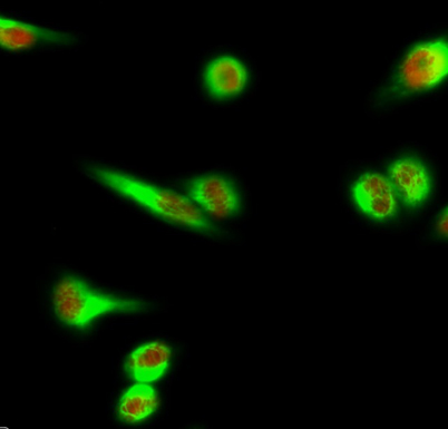 Immunofluorescence analysis of Hela cell. 1,Lamin A/C Polyclonal Antibody(red) was diluted at 1:200(4° overnight). Galectin-3 Monoclonal Antibody(6G2)(green) was diluted at 1:200(4° overnight). 2, Goat Anti Rabbit Alexa Fluor 594 Catalog:RS3611 was dilute
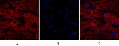 Immunofluorescence analysis of human-liver tissue. 1,Lamin A/C Polyclonal Antibody(red) was diluted at 1:200(4°C,overnight). 2, Cy3 labled Secondary antibody was diluted at 1:300(room temperature, 50min).3, Picture B: DAPI(blue) 10min. Picture A:Target. P
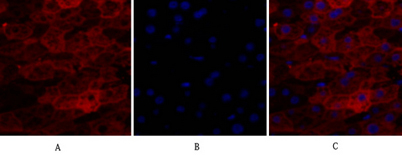 Immunofluorescence analysis of human-liver tissue. 1,Lamin A/C Polyclonal Antibody(red) was diluted at 1:200(4°C,overnight). 2, Cy3 labled Secondary antibody was diluted at 1:300(room temperature, 50min).3, Picture B: DAPI(blue) 10min. Picture A:Target. P
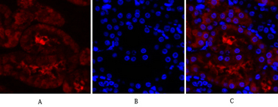 Immunofluorescence analysis of rat-kidney tissue. 1,Lamin A/C Polyclonal Antibody(red) was diluted at 1:200(4°C,overnight). 2, Cy3 labled Secondary antibody was diluted at 1:300(room temperature, 50min).3, Picture B: DAPI(blue) 10min. Picture A:Target. Pi