
ELK Biotechnology
SKU:ES2625
InsP 3-kinase C Rabbit Polyclonal Antibody
InsP 3-kinase C Rabbit Polyclonal Antibody
Regular price
$248.00 USD
Regular price
Sale price
$248.00 USD
Unit price
per
Shipping calculated at checkout.
Couldn't load pickup availability
InsP 3-kinase C Rabbit Polyclonal Antibody
Overview
-
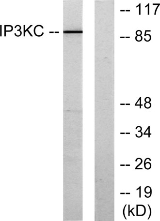
 Western Blot analysis of various cells using InsP 3-kinase C Polyclonal Antibody diluted at 1:2000
 -
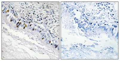 Immunohistochemical analysis of paraffin-embedded Human lung cancer. Antibody was diluted at 1:100(4° overnight). High-pressure and temperature Tris-EDTA,pH8.0 was used for antigen retrieval. Negetive contrl (right) obtaned from antibody was pre-absorbe
 -
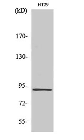
 Western blot analysis of lysates from HT-29 cells, using IP3KC Antibody. The lane on the right is blocked with the synthesized peptide.
