IK; RED; RER; Protein Red; Cytokine IK; IK factor; Protein RER
Applications:
WB;IHC;IF;ELISA
Recommended Dilutions:
Western Blot: 1/500 - 1/2000. Immunohistochemistry: 1/100 - 1/300. ELISA: 1/20000. Not yet tested in other applications.
Immunogen:
The antiserum was produced against synthesized peptide derived from human RED. AA range:508-557
Storage:
Rabbit
Storage:
-20°C/1 year
Clonality:
Polyclonal
Isotype:
IgG
Concentration:
1 mg/ml
Observed Band:
66kD
GeneID(Human):
3550
Human Swiss-Prot No:
Q13123
Cellular localization:
Nucleus . Nucleus, nucleoplasm . Chromosome . Cytoplasm, cytoskeleton, spindle pole . Predominantly present throughout the nucleoplasm during prometaphase, metaphase and anaphase. Is also detected in nuclear foci that are not identical with Cajal bodies. Starts to accumulate at chromosomes during telophase, and is nearly exclusively associated with chromosomes in newly divided cells (PubMed:24252166). Colocalizes with MAD1L1 at mitotic spindle poles during metaphase and anaphase (PubMed:22351768). .
Background:
The protein encoded by this gene was identified by its RED repeat, a stretch of repeated arginine, glutamic acid and aspartic acid residues. The protein localizes to discrete dots within the nucleus, excluding the nucleolus. Its function is unknown. This gene maps to chromosome 5; however, a pseudogene may exist on chromosome 2. [provided by RefSeq, Jul 2008],

 Western Blot analysis of various cells using IK Polyclonal Antibody diluted at 1:2000 cells nucleus extracted by Minute TM Cytoplasmic and Nuclear Fractionation kit (SC-003,Inventbiotech,MN,USA).
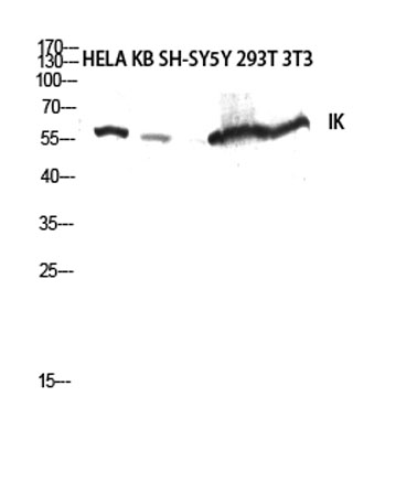 Western blot analysis of HELA KB SH-SY5Y 293T 3T3 lysis using IK antibody. Antibody was diluted at 1:2000 cells nucleus extracted by Minute TM Cytoplasmic and Nuclear Fractionation kit (SC-003,Inventbiotech,MN,USA).
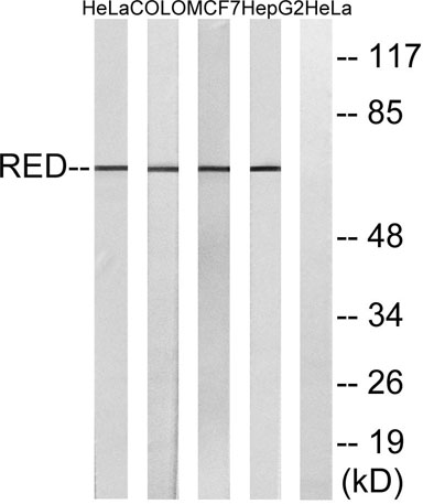 Western blot analysis of lysates from HepG2, MCF-7, COLO, and HeLa cells, using RED Antibody. The lane on the right is blocked with the synthesized peptide.
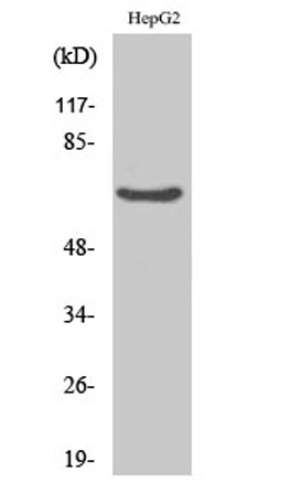
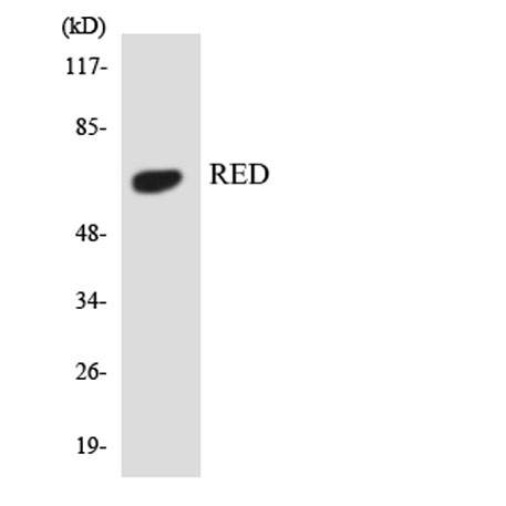 Western blot analysis of the lysates from 293 cells using RED antibody.