ELK Biotechnology
SKU:ES2383
G2A Rabbit Polyclonal Antibody
G2A Rabbit Polyclonal Antibody
Regular price
$248.00 USD
Regular price
Sale price
$248.00 USD
Unit price
per
Shipping calculated at checkout.
Couldn't load pickup availability
G2A Rabbit Polyclonal Antibody
Overview
-
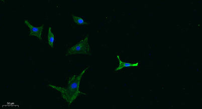 Immunofluorescence analysis of A549. 1,primary Antibody was diluted at 1:200(4°C overnight). 2, Goat Anti Rabbit IgG (H&L) - Alexa Fluor 488 Secondary antibody was diluted at 1:1000(room temperature, 50min).3, Picture B: DAPI(blue) 10min.
 -
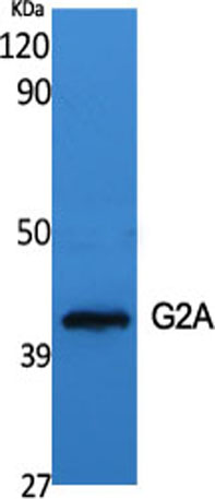 Western Blot analysis of various cells using G2A Polyclonal Antibody diluted at 1:500
 -
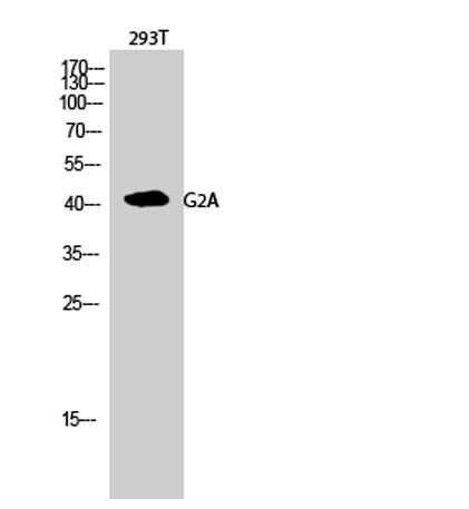 Western Blot analysis of 293T cells using G2A Polyclonal Antibody diluted at 1:500
 -
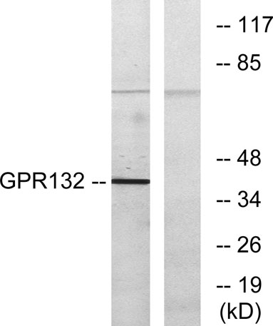 Western blot analysis of lysates from COS7 cells, using GPR132 Antibody. The lane on the right is blocked with the synthesized peptide.