Western Blot: 1/500 - 1/2000. Immunohistochemistry: 1/100 - 1/300. Immunofluorescence: 1/200 - 1/1000. ELISA: 1/40000. Not yet tested in other applications.
Immunogen:
The antiserum was produced against synthesized peptide derived from human FAS ligand. AA range:101-150
Storage:
Rabbit
Storage:
-20°C/1 year
Clonality:
Polyclonal
Isotype:
IgG
Concentration:
1 mg/ml
Observed Band:
33kD
GeneID(Human):
356
Human Swiss-Prot No:
P48023
Cellular localization:
Cell membrane ; Single-pass type II membrane protein . Cytoplasmic vesicle lumen . Lysosome lumen . Is internalized into multivesicular bodies of secretory lysosomes after phosphorylation by FGR and monoubiquitination (PubMed:17164290). Colocalizes with the SPPL2A protease at the cell membrane (PubMed:17557115). .; [Tumor necrosis factor ligand superfamily member 6, soluble form]: Secreted . May be released into the extracellular fluid by cleavage from the cell surface. .; [FasL intracellular domain]: Nucleus . The FasL ICD cytoplasmic form is translocated into the nucleus. .
Background:
This gene is a member of the tumor necrosis factor superfamily. The primary function of the encoded transmembrane protein is the induction of apoptosis triggered by binding to FAS. The FAS/FASLG signaling pathway is essential for immune system regulation, including activation-induced cell death (AICD) of T cells and cytotoxic T lymphocyte induced cell death. It has also been implicated in the progression of several cancers. Defects in this gene may be related to some cases of systemic lupus erythematosus (SLE). Alternatively spliced transcript variants have been described. [provided by RefSeq, Nov 2014],

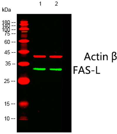 Western blot analysis of lysates from 1)HepG2, 2)293 cells, (Green) primary antibody was diluted at 1:1000, 4°over night, Dylight 800 secondary antibody(Immunoway:RS23920)was diluted at 1:10000, 37° 1hour. (Red) Actin β Monoclonal Antibody(5G3) (Immunoway
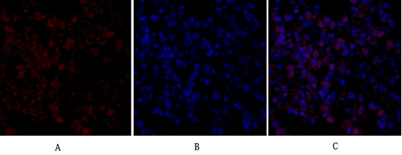 Immunofluorescence analysis of rat-lung tissue. 1,FAS-L Polyclonal Antibody(red) was diluted at 1:200(4°C,overnight). 2, Cy3 labled Secondary antibody was diluted at 1:300(room temperature, 50min).3, Picture B: DAPI(blue) 10min. Picture A:Target. Picture
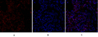 Immunofluorescence analysis of rat-lung tissue. 1,FAS-L Polyclonal Antibody(red) was diluted at 1:200(4°C,overnight). 2, Cy3 labled Secondary antibody was diluted at 1:300(room temperature, 50min).3, Picture B: DAPI(blue) 10min. Picture A:Target. Picture
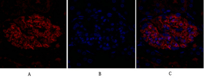 Immunofluorescence analysis of rat-kidney tissue. 1,FAS-L Polyclonal Antibody(red) was diluted at 1:200(4°C,overnight). 2, Cy3 labled Secondary antibody was diluted at 1:300(room temperature, 50min).3, Picture B: DAPI(blue) 10min. Picture A:Target. Pictur