ELK Biotechnology
SKU:ES2210
Dyrk1A Rabbit Polyclonal Antibody
Dyrk1A Rabbit Polyclonal Antibody
Regular price
$248.00 USD
Regular price
Sale price
$248.00 USD
Unit price
per
Shipping calculated at checkout.
Couldn't load pickup availability
Dyrk1A Rabbit Polyclonal Antibody
Overview
-
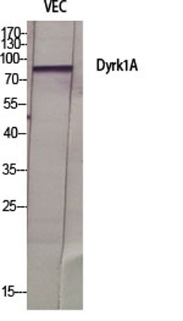 Western Blot analysis of various cells using Dyrk1A Polyclonal Antibody diluted at 1:500 cells nucleus extracted by Minute TM Cytoplasmic and Nuclear Fractionation kit (SC-003,Inventbiotech,MN,USA).
 -
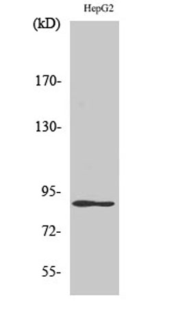 Western Blot analysis of HepG2 cells using Dyrk1A Polyclonal Antibody diluted at 1:500 cells nucleus extracted by Minute TM Cytoplasmic and Nuclear Fractionation kit (SC-003,Inventbiotech,MN,USA).
 -
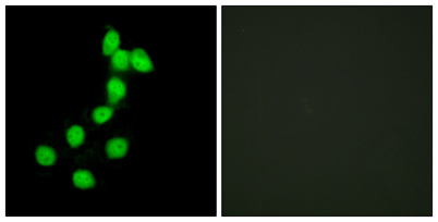 Immunofluorescence analysis of HepG2 cells, using DYR1A Antibody. The picture on the right is blocked with the synthesized peptide.
 -
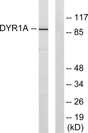 Western blot analysis of lysates from HepG2 cells, using DYR1A Antibody. The lane on the right is blocked with the synthesized peptide.