
ELK Biotechnology
SKU:ES2172
Dio-1 Rabbit Polyclonal Antibody
Dio-1 Rabbit Polyclonal Antibody
Regular price
$248.00 USD
Regular price
Sale price
$248.00 USD
Unit price
per
Shipping calculated at checkout.
Couldn't load pickup availability
Dio-1 Rabbit Polyclonal Antibody
Overview
-
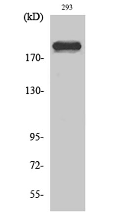 Western Blot analysis of various cells using Dio-1 Polyclonal Antibody diluted at 1:2000
 -
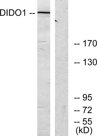 Western blot analysis of lysates from 293 cells, using DIDO1 Antibody. The lane on the right is blocked with the synthesized peptide.
 -
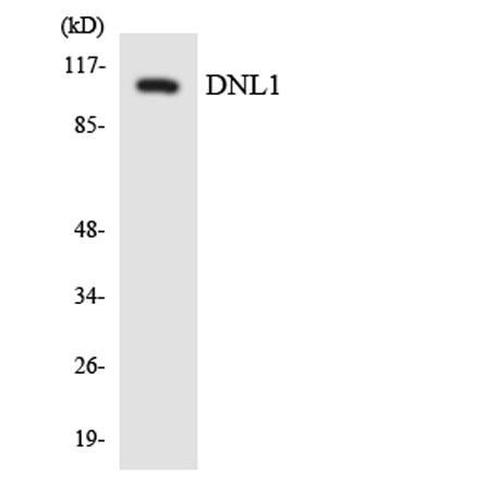 Western blot analysis of the lysates from HepG2 cells using DNL1 antibody.
 -
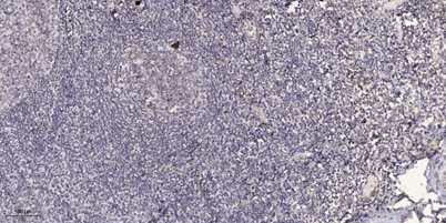 Immunohistochemical analysis of paraffin-embedded human tonsil. 1, Antibody was diluted at 1:200(4° overnight). 2, Tris-EDTA,pH9.0 was used for antigen retrieval. 3,Secondary antibody was diluted at 1:200(room temperature, 45min).
