ATXN2; ATX2; SCA2; TNRC13; Ataxin-2; Spinocerebellar ataxia type 2 protein; Trinucleotide repeat-containing gene 13 protein
Applications:
WB;IHC;IF;ELISA
Recommended Dilutions:
Western Blot: 1/500 - 1/2000. Immunohistochemistry: 1/100 - 1/300. ELISA: 1/20000. Not yet tested in other applications.
Immunogen:
The antiserum was produced against synthesized peptide derived from human ATXN2. AA range:731-780
Storage:
-20°C/1 year
Storage:
Rabbit
Clonality:
Polyclonal
Isotype:
IgG
Concentration:
1 mg/ml
Observed Band:
140kD
GeneID(Human):
6311
Human Swiss-Prot No:
Q99700
Cellular localization:
Cytoplasm .
Background:
ataxin 2(ATXN2) Homo sapiens This gene belongs to a group of genes that is associated with microsatellite-expansion diseases, a class of neurological and neuromuscular disorders caused by expansion of short stretches of repetitive DNA. The protein encoded by this gene has two globular domains near the N-terminus, one of which contains a clathrin-mediated trans-Golgi signal and an endoplasmic reticulum exit signal. The protein is primarily localized to the Golgi apparatus, with deletion of the Golgi and endoplasmic reticulum signals resulting in abnormal subcellular localization. In addition, the N-terminal region contains a polyglutamine tract of 14-31 residues that can be expanded in the pathogenic state to 32-200 residues. Intermediate length expansions of this tract increase susceptibility to amyotrophic lateral sclerosis, while long expansions of this tract result in spinocerebellar ataxia-2, an autosomal-dominantly inherited, neurodegener

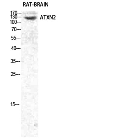 Western Blot analysis of various cells using Ataxin-2 Polyclonal Antibody diluted at 1:1000
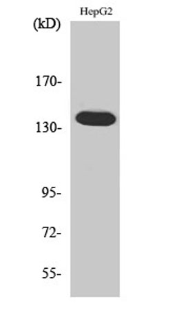 Western Blot analysis of 293 cells using Ataxin-2 Polyclonal Antibody diluted at 1:1000
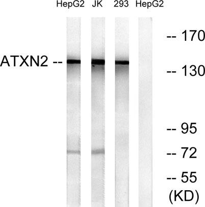 Western blot analysis of lysates from HepG2, Jurkat, and 293 cells, using ATXN2 Antibody. The lane on the right is blocked with the synthesized peptide.
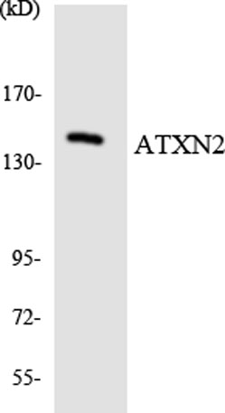 Western blot analysis of the lysates from HepG2 cells using ATXN2 antibody.