ATXN1; ATX1; SCA1; Ataxin-1; Spinocerebellar ataxia type 1 protein
Applications:
WB;IHC;IF;ELISA
Recommended Dilutions:
Immunohistochemistry: 1/100 - 1/300. Immunofluorescence: 1/200 - 1/1000. ELISA: 1/5000. Not yet tested in other applications.
Immunogen:
The antiserum was produced against synthesized peptide derived from human Ataxin 1. AA range:742-791
Storage:
Rabbit
Storage:
-20°C/1 year
Clonality:
Polyclonal
Isotype:
IgG
Concentration:
1 mg/ml
Observed Band:
87kD
GeneID(Human):
6310
Human Swiss-Prot No:
P54253
Cellular localization:
Cytoplasm . Nucleus . Colocalizes with USP7 in the nucleus. .
Background:
ataxin 1(ATXN1) Homo sapiens The autosomal dominant cerebellar ataxias (ADCA) are a heterogeneous group of neurodegenerative disorders characterized by progressive degeneration of the cerebellum, brain stem and spinal cord. Clinically, ADCA has been divided into three groups: ADCA types I-III. ADCAI is genetically heterogeneous, with five genetic loci, designated spinocerebellar ataxia (SCA) 1, 2, 3, 4 and 6, being assigned to five different chromosomes. ADCAII, which always presents with retinal degeneration (SCA7), and ADCAIII often referred to as the `pure' cerebellar syndrome (SCA5), are most likely homogeneous disorders. Several SCA genes have been cloned and shown to contain CAG repeats in their coding regions. ADCA is caused by the expansion of the CAG repeats, producing an elongated polyglutamine tract in the corresponding protein. The expanded repeats are variable in size and unstable, usually increasing in size when transmitted

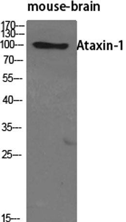 Western Blot analysis of various cells using Ataxin-1 Polyclonal Antibody diluted at 1:500
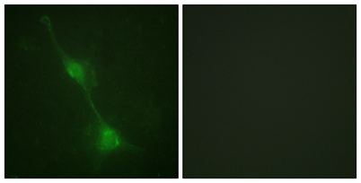 Immunofluorescence analysis of NIH/3T3 cells, using Ataxin 1 Antibody. The picture on the right is blocked with the synthesized peptide.
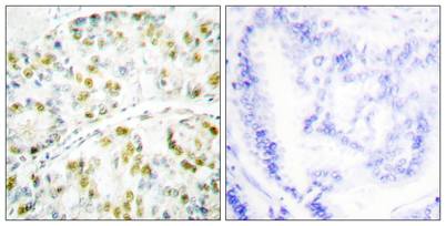 Immunohistochemistry analysis of paraffin-embedded human lung carcinoma tissue, using Ataxin 1 Antibody. The picture on the right is blocked with the synthesized peptide.