ELK Biotechnology
SKU:ES1648
AMPKα1 Rabbit Polyclonal Antibody
AMPKα1 Rabbit Polyclonal Antibody
Regular price
$248.00 USD
Regular price
Sale price
$248.00 USD
Unit price
per
Shipping calculated at checkout.
Couldn't load pickup availability
AMPKα1 Rabbit Polyclonal Antibody
Overview
-
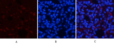 Immunofluorescence analysis of rat-lung tissue. 1,AMPKα1 Polyclonal Antibody(red) was diluted at 1:200(4°C,overnight). 2, Cy3 labled Secondary antibody was diluted at 1:300(room temperature, 50min).3, Picture B: DAPI(blue) 10min. Picture A:Target. Picture
 -
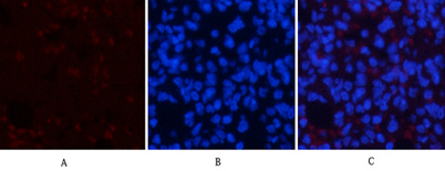 Immunofluorescence analysis of rat-lung tissue. 1,AMPKα1 Polyclonal Antibody(red) was diluted at 1:200(4°C,overnight). 2, Cy3 labled Secondary antibody was diluted at 1:300(room temperature, 50min).3, Picture B: DAPI(blue) 10min. Picture A:Target. Picture
 -
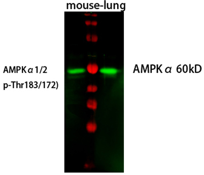 Western Blot analysis of mouse-lung cells using primary antibody diluted at 1:1000(4°C overnight). Secondary antibody:Goat Anti-rabbit IgG IRDye 800( diluted at 1:5000, 25°C, 1 hour). Cell lysate was extracted by Minute™ Plasma Membrane Protein Isolation
 -
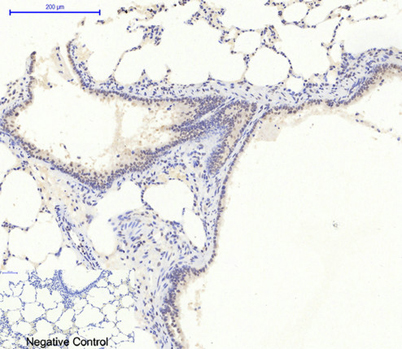 Immunohistochemical analysis of paraffin-embedded Rat-lung tissue. 1,AMPKα1 Polyclonal Antibody was diluted at 1:200(4°C,overnight). 2, Sodium citrate pH 6.0 was used for antibody retrieval(>98°C,20min). 3,Secondary antibody was diluted at 1:200(room temp