1
/
of
1
ELK Biotechnology
SKU:ES2327
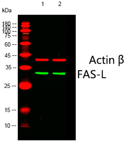
FAS-L rabbit pAb
FAS-L rabbit pAb
Regular price
$250.00 USD
Regular price
Sale price
$250.00 USD
Unit price
/
per
Shipping calculated at checkout.
Couldn't load pickup availability
Applications: WB;IHC;IF;ELISA
Reactivity: Human;Mouse;Pig
Source: Rabbit
Dilution: Western Blot: 1/500 - 1/2000. Immunohistochemistry: 1/100 - 1/300. Immunofluorescence: 1/200 - 1/1000. ELISA: 1/40000. Not yet tested in other applications.
Immunogen: The antiserum was produced against synthesized peptide derived from human FAS ligand. AA range:101-150
Storage_stability: -20°C/1 year
Clonality: Polyclonal
Isotype: IgG
Concentration: 1 mg/ml
Observed_band(KD): 33kD
Human_gene_id: 356
Human_swiss_prot_no: P48023
Subcellular_location: Cell membrane ; Single-pass type II membrane protein . Cytoplasmic vesicle lumen . Lysosome lumen . Is internalized into multivesicular bodies of secretory lysosomes after phosphorylation by FGR and monoubiquitination (PubMed:17164290). Colocalizes with the SPPL2A protease at the cell membrane (PubMed:17557115). .; [Tumor necrosis factor ligand superfamily member 6, soluble form]: Secreted . May be released into the extracellular fluid by cleavage from the cell surface. .; [FasL intracellular domain]: Nucleus . The FasL ICD cytoplasmic form is translocated into the nucleus. .
Other_name: FASLG; APT1LG1; CD95L; FASL; TNFSF6; Tumor necrosis factor ligand superfamily member 6; Apoptosis antigen ligand; APTL; CD95 ligand; CD95-L; Fas antigen ligand; Fas ligand; FasL; CD antigen CD178
Background: This gene is a member of the tumor necrosis factor superfamily. The primary function of the encoded transmembrane protein is the induction of apoptosis triggered by binding to FAS. The FAS/FASLG signaling pathway is essential for immune system regulation, including activation-induced cell death (AICD) of T cells and cytotoxic T lymphocyte induced cell death. It has also been implicated in the progression of several cancers. Defects in this gene may be related to some cases of systemic lupus erythematosus (SLE). Alternatively spliced transcript variants have been described. [provided by RefSeq, Nov 2014],
Share