1
/
of
1
ELK Biotechnology
SKU:ES14389
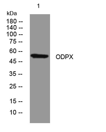
ODPX rabbit pAb
ODPX rabbit pAb
Regular price
$250.00 USD
Regular price
Sale price
$250.00 USD
Unit price
/
per
Shipping calculated at checkout.
Couldn't load pickup availability
Applications: WB
Reactivity: Human; Mouse
Source: Rabbit
Dilution: WB 1:500-2000
Immunogen: Synthesized peptide derived from human ODPX AA range: 24-74
Storage_stability: -20°C/1 year
Clonality: Polyclonal
Isotype: IgG
Concentration: 1 mg/ml
Human_gene_id: 8050
Human_swiss_prot_no: O00330
Subcellular_location: Mitochondrion matrix.
Background: The pyruvate dehydrogenase (PDH) complex is located in the mitochondrial matrix and catalyzes the conversion of pyruvate to acetyl coenzyme A. The PDH complex thereby links glycolysis to Krebs cycle. The PDH complex contains three catalytic subunits, E1, E2, and E3, two regulatory subunits, E1 kinase and E1 phosphatase, and a non-catalytic subunit, E3 binding protein (E3BP). This gene encodes the E3 binding protein subunit; also known as component X of the pyruvate dehydrogenase complex. This protein tethers E3 dimers to the E2 core of the PDH complex. Defects in this gene are a cause of pyruvate dehydrogenase deficiency which results in neurological dysfunction and lactic acidosis in infancy and early childhood. This protein is also a minor antigen for antimitochondrial antibodies. These autoantibodies are present in nearly 95% of patients with the autoimmune liver disease primary biliary cirrhosis (PBC). In PBC, activated T lymphocytes attack and destroy epithelial cells in the bile duct where this protein is abnormally distributed and overexpressed. PBC eventually leads to cirrhosis and liver failure. Alternative splicing results in multiple transcript variants encoding distinct isoforms.[provided by RefSeq, Oct 2009],
Share