1
/
of
1
ELK Biotechnology
SKU:EM1336
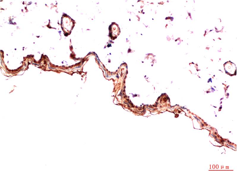
PDGFRα (5D1) Mouse mAb
PDGFRα (5D1) Mouse mAb
Regular price
$250.00 USD
Regular price
Sale price
$250.00 USD
Unit price
/
per
Shipping calculated at checkout.
Couldn't load pickup availability
Applications: IHC
Reactivity: Human, Mouse, Rat
Source: Mouse
Dilution: IHC 1:100-200
Immunogen: Synthetic Peptide
Storage_stability: PBS with 0.02% sodium azide and 50% glycerol pH 7.4. Store at -20°C. Avoid repeated freeze-thaw cycles.
Clonality: Monoclonal
Isotype: IgG1
Concentration: 1mg/mL
Observed_band(KD): 180kDa
Human_gene_id: 5156
Human_swiss_prot_no: P16234
Subcellular_location: Membrane
Background: Platelet derived growth factor (PDGF) family proteins exist as several disulphide-bonded, dimeric isoforms (PDGF AA, PDGF AB, PDGF BB, PDGF CC, and PDGF DD) that bind in a specific pattern to two closely related receptor tyrosine kinases, PDGF receptor α (PDGFRα) and PDGF receptor β (PDGFRβ). PDGFRα and PDGFRβ can each form heterodimers with EGFR, which is also activated by PDGF. Various cells differ in the total number of receptors present and in the receptor subunit composition, which may account for responsive differences among cell types to PDGF binding. Ligand binding induces receptor dimerization and autophosphorylation, followed by binding and activation of cytoplasmic SH2 domain-containing signal transduction molecules, such as GRB2, Src, GAP, PI3 kinase, PLCγ, and NCK. A number of different signaling pathways are initiated by activated PDGF receptors and lead to control of cell growth, actin reorganization, migration, and differentiation.
Share