

Cusabio
SKU:CSB-RA043670A0HU
U2AF2 Recombinant Monoclonal Antibody
U2AF2 Recombinant Monoclonal Antibody
Couldn't load pickup availability
| Release Date | 2025-10-28 |
|---|---|
| Product Code | CSB-RA043670A0HU |
| Short Name | U2AF2 Recombinant Monoclonal Antibody |
| Antibody Type | Recombinant Antibody |
| List Price | $350/100ul, $210/50ul |
| Species Reactivity | Human, Mouse, Rat |
| Tested Applications | ELISA, WB, IHC, IF, FC |
| Dilution | WB:1:500-1:5000, IHC:1:50-1:200, IF:1:50-1:200, FC:1:50-1:200 |
| Purification Method | Affinity chromatography |
| Buffer Formulation | PBS, 150mM NaCl, 50% Glycerol, 0.05% BSA, 0.02% Sodium Azide, pH 7.4. |
| Antigen | A synthesized peptide from human U2AF2 protein |
| Antigen Species | Human |
| Uniprot ID | P26368 |
| Target Name | U2AF2 |
| Clonality | Monoclonal |
| Clone ID | 1H1 |
| Host Species | Rabbit |
| Isotype | IgG |
| Conjugation | Non-conjugated |
| Form | Liquid |
| Storage Condition | Upon delivery aliquot and store at -20°C or -80°C. Avoid repeated freeze. |
| TDS URL | https://www.cusabio.com/datasheet/12946172/ |
| Size | 50ul |
Image 1
Western Blot
,
Positive WB detected in:HEK293 whole cell lysate(30µg), HT29 whole cell lysate(30µg), Jurkat whole cell lysate(30µg), HepG2 whole cell lysate(30µg), A431 whole cell lysate(30µg), A549 whole cell lysate(30µg), Mouse brain tissue lysate(30µg), Rat brain lysate(30µg)
All lanes: U2AF2 antibody at 1:1000
Secondary
Goat polyclonal to rabbit IgG at 1/40000 dilution
Predicted band size: 54 kDa
Observed band size: 60 kDa
Exposure time:20s
Image 2
IHC image of CSB-RA043670A0HU diluted at 1:100 and staining in paraffin-embedded human colorectal cancer performed on a Leica BondTM system. After dewaxing and hydration, antigen retrieval was mediated by high pressure in a citrate buffer (pH 6.0). Section was blocked with 10% normal goat serum 30min at RT. Then primary antibody (1% BSA) was incubated at 4°C overnight. The primary is detected by a Goat anti-rabbit polymer IgG labeled by HRP and visualized using 0.05% DAB.
Image 3
Immunofluorescence staining of A-431 cell with CSB-RA043670A0HU at 1:50 , counter-stained with DAPI. The cells were fixed in 4% formaldehyde, permeabilized using 0.2% Triton X-100 and blocked in 10% normal Goat Serum. The cells were then incubated with the antibody overnight at 4°C. The secondary antibody was Alexa Fluor 488-congugated AffiniPure Goat Anti-Rabbit IgG(H+L).
Image 4
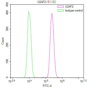
Overlay Peak curve showing Hela cells stained with CSB-RA043670A0HU (red line) at 1:100. The cells were fixed in 4% formaldehyde and permeated by 0.2% TritonX-100 for 10min. Then 10% normal goat serum to block non-specific protein-protein interactions followed by the antibody (1ug/1*10<sup>6</sup>cells) for 45min at 4℃. The secondary antibody used was FITC-conjugated goat anti-rabbit IgG (H+L) at 1/200 dilution for 35min at 4℃.Control antibody (green line) was Rabbit IgG (1ug/1*10<sup>6</sup>cells) used under the same conditions. Acquisition of >10, 000 events was performed.
Share