

Cusabio
SKU:CSB-RA006255MA1HU
CXCR5 Recombinant Monoclonal Antibody
CXCR5 Recombinant Monoclonal Antibody
Couldn't load pickup availability
| Release Date | 2025-10-16 |
|---|---|
| Product Code | CSB-RA006255MA1HU |
| Short Name | CXCR5 Recombinant Monoclonal Antibody |
| Antibody Type | Recombinant Antibody |
| List Price | $350/100ul, $210/50ul |
| Species Reactivity | Human |
| Tested Applications | ELISA, WB, IHC, IF, FC |
| Dilution | WB:1:500-1:2000, IHC:1:50-1:200, IF:1:50-1:200, FC:1:50-1:200 |
| Purification Method | Affinity chromatography |
| Buffer Formulation | 0.03% Proclin 300, 50% Glycerol, 0.01M PBS, pH 7.4. |
| Antigen | Recombinant Human CXCR5 protein |
| Antigen Species | Human |
| Uniprot ID | P32302 |
| Target Name | CXCR5 |
| Clonality | Monoclonal |
| Clone ID | 25A3 |
| Host Species | Human |
| Isotype | IgG1 |
| Conjugation | Non-conjugated |
| Form | Liquid |
| Storage Condition | Upon delivery aliquot and store at -20°C or -80°C. Avoid repeated freeze. |
| TDS URL | https://www.cusabio.com/datasheet/12945807/ |
| Size | 100ul |
Image 1
Western Blot
Positive WB detected in: Jurkat whole cell lysate(30µg), Raji whole cell lysate(30µg), SH-SY5Y whole cell lysate(30µg), U-87MG whole cell lysate(30µg), CT26 whole cell lysate(30µg), PC-3 whole cell lysate(30µg), MCF-7 whole cell lysate(30µg), HL-60 whole cell lysate(30µg)
All lanes: CXCR5 antibody at 1:1000
Secondary
Goat polyclonal to human IgG at 1/40000 dilution
Predicted band size: 42 kDa
Observed band size: 33 kDa
Exposure time: 2min
Image 2
IHC image of CSB-RA006255MA1HU diluted at 1:50 and staining in paraffin-embedded human lymph node tissue performed on a Leica BondTM system. After dewaxing and hydration, antigen retrieval was mediated by high pressure in a citrate buffer (pH 6.0). Section was blocked with 10% normal goat serum 30min at RT. Then primary antibody (1% BSA) was incubated at 4°C overnight. The primary is detected by a Anti-Human lgG, Fcy Fragment Specific labeled by HRP and visualized using 0.05% DAB.
Image 3
Immunofluorescence staining of HepG2 cell with CSB-RA006255MA1HU at 1:30 ,counter-stained with DAPI. The cells were fixed in 4% formaldehyde and blocked in 10% normal Goat Serum. The cells were then incubated with the antibody overnight at 4°C. The secondary antibody was Fluorescein (FITC) AffiniPure Goat Anti-Human IgG, Fcγ fragment specific.
Image 4
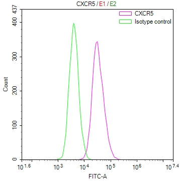
Overlay Peak curve showing Raji cells stained with CSB-RA006255MA1HU (red line) at 1:100. Then 10% normal goat serum to block non-specific protein-protein interactions followed by the antibody (1ug/1*10<sup>6</sup>cells) for 45min at 4℃. The secondary antibody used was Fluorescein (FITC) AffiniPure Goat Anti-Human IgG, Fcγ fragment specific at 1:200 dilution for 35min at 4℃.Control antibody (green line) was human IgG1 (1ug/1*10<sup>6</sup>cells) used under the same conditions. Acquisition of >10,000 events was performed.
Share