

Cusabio
SKU:CSB-PA012742LA01HU
LANCL1 Antibody
LANCL1 Antibody
Couldn't load pickup availability
| Release Date | 2022-04-01 |
|---|---|
| Product Code | CSB-PA012742LA01HU |
| Short Name | LANCL1 Antibody |
| Antibody Type | Polyclonal Antibody |
| List Price | $299/100ug, $166/50ug |
| Species Reactivity | Human, Mouse, Rat |
| Tested Applications | ELISA, WB, IHC |
| Dilution | WB:1:1000-1:5000, IHC:1:200-1:500 |
| Purification Method | Antigen affinity purification |
| Buffer Formulation | PBS, 50% Glycerol, 0.5% BSA, 0.02% Sodium Azide, pH 7.4. |
| Antigen | Synthesized peptide derived from human LANCL1 |
| Antigen Species | Human |
| Uniprot ID | O43813 |
| Target Name | LANCL1 |
| Clonality | Polyclonal |
| Host Species | Rabbit |
| Isotype | IgG |
| Conjugation | Non-conjugated |
| Form | Liquid |
| Storage Condition | Upon delivery aliquot and store at -20°C or -80°C. Avoid repeated freeze. |
| TDS URL | https://www.cusabio.com/datasheet/12549058/ |
| Size | 100ug |
Image 1
Western Blot
Positive WB detected in: Mouse Brain tissue lysate, Mouse Kidney tissue lysate, Mouse skeletal muscle tissue lysate, Mous Liver tissue lysate, Rat Kidney tissue lysate, Rat Heart tissue lysate, A549 whole cell lysate, Hela whole cell lysate, HepG2 whole cell lysate
All lanes: LANCL1 antibody at 1:1000
Secondary
Goat polyclonal to rabbit IgG at 1/50000 dilution
Predicted band size: 42 kDa
Observed band size: 42 kDa
Image 2
IHC image of CSB-PA012742LA01HU diluted at 1:300 and staining in paraffin-embedded human testis tissue performed on a Leica BondTM system. After dewaxing and hydration, antigen retrieval was mediated by high pressure in a citrate buffer (pH 6.0). Section was blocked with 10% normal goat serum 30min at RT. Then primary antibody (1% BSA) was incubated at 4°C overnight. The primary is detected by a Goat anti-rabbit polymer IgG labeled by HRP and visualized using 0.05% DAB.
Secondary antibody only control: uses 1% BSA instead of primary antibody
Image 3
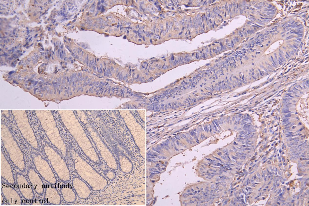
IHC image of CSB-PA012742LA01HU diluted at 1:300 and staining in paraffin-embedded human colorectal cancer performed on a Leica BondTM system. After dewaxing and hydration, antigen retrieval was mediated by high pressure in a citrate buffer (pH 6.0). Section was blocked with 10% normal goat serum 30min at RT. Then primary antibody (1% BSA) was incubated at 4°C overnight. The primary is detected by a Goat anti-rabbit polymer IgG labeled by HRP and visualized using 0.05% DAB.
Secondary antibody only control: uses 1% BSA instead of primary antibody
Share